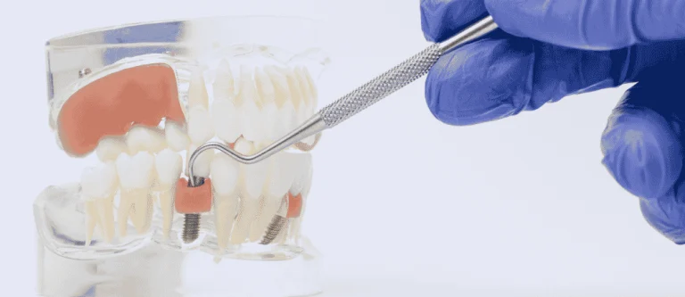
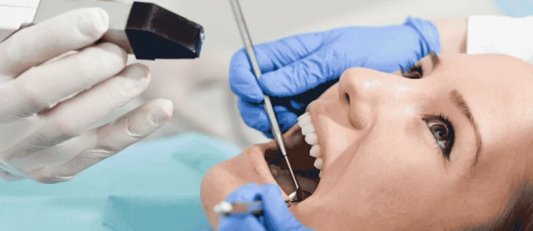
When you plan for dental implants, you need strong bone to hold them in place. Your dentist does not guess about your bone density.
They use clear tests and images to measure the strength and thickness of your jaw before implant surgery.
Dentists measure bone density before implant surgery by using 3D scans like cone beam computed tomography (CBCT), digital imaging, and clinical exams to check bone quality, volume, and shape.
These tools show how much bone you have and how dense it is. This information helps your dentist choose the right implant size and position to improve implant success.
If your bone looks thin or soft, your dentist can adjust the treatment plan. You may need bone grafting or other support before placing the implant.
When you understand how implant dentistry measures bone density, you can feel more confident about your care.
Key Takeaways
- Dentists use 3D scans and exams to measure bone density before placing dental implants.
- Bone quality and volume directly affect implant success and stability.
- Your treatment plan may change if you have low bone density.
Why Bone Density Matters for Implant Outcomes
Bone density directly affects how stable your dental implant feels on the day of surgery and how well it bonds to your jaw over time.
The strength and structure of your jawbone shape implant stability, healing speed, and long‑term dental implant success.
Relationship Between Bone Quality and Implant Stability
Your dentist looks at both bone density and bone quality before placing dental implants. Dense bone, especially strong cortical bone, grips the implant threads more tightly.
This creates better primary stability, also called initial stability, right after placement.
Lower-density bone often contains more cancellous bone density and less thick cortical bone. This softer structure may not hold the implant as firmly at first.
When initial stability is weak, the risk of movement increases, and movement can disrupt healing.
Studies explain that bone density plays a key role in primary stability and implant success. Higher jawbone density usually means stronger mechanical support. Lower density may require changes in implant size, drilling method, or healing time.
In simple terms, the stronger your jawbone density, the better your implant can lock into place from the start.
Impact on Osseointegration and Healing
After placement, your implant relies on osseointegration. This process allows bone cells to grow and attach directly to the implant surface. Good bone density supports faster and more predictable bone healing.
When your bone is dense, it often provides a stable base for new bone growth. This can shorten healing time and improve long‑term dental implant success.
Strong bone also reduces the chance of tiny movements that interrupt osseointegration.
Cone beam scans help measure jawbone density before surgery. Dentists commonly use CBCT imaging to evaluate cancellous bone density. These scans guide decisions about implant length, width, and placement angle.
If your bone density is lower, healing may take longer. Your dentist may suggest staged treatment or bone grafting to improve the site before implant placement.
Consequences of Low Bone Density and Implant Failure
Low jawbone density increases the risk of implant failure, especially in the early stages. If the implant lacks primary stability, small forces from chewing can cause micro-movements. These movements can prevent proper osseointegration.
Poor bone quality may also increase the chance of bone resorption around the implant. As bone resorbs, the implant loses support. Over time, this can lead to looseness or complete failure.
Research on bone density requirements for dental implants shows that adequate density improves stability and long‑term outcomes. When density falls below ideal levels, your dentist may adjust the surgical plan.
Options include wider implants, deeper placement into cortical bone, or bone graft procedures.
By understanding your bone density, you and your dentist can reduce risks and improve the odds of lasting implant success.
Diagnostic Tools for Assessing Jawbone Health
Dentists rely on detailed imaging to check your jawbone before implant surgery. These tools show bone height, width, and density so your dentist can plan safe and stable implant placement.

CBCT and Cone Beam Computed Tomography
CBCT (cone beam computed tomography) gives your dentist a 3D view of your jaw. Unlike standard dental X-rays, CBCT scans capture thin slices of bone and combine them into a detailed 3D image.
With dental cone beam computed tomography (CBCT), your dentist can examine the exact implant site. They can measure bone width, height, and the quality of cancellous bone inside the jaw.
This matters because implants need enough strong bone to stay stable.
CBCT also helps your dentist avoid important structures. These include nerves, sinus cavities, and nearby tooth roots. The scan supports precise bone density measurement by analyzing grayscale values within specific regions of interest.
Micro-CT can measure bone in even finer detail, but dentists mainly use it for research, not routine patient care.
Role of Panoramic X-Rays and 3D Imaging
A panoramic X-ray shows your entire upper and lower jaw in one flat image. It gives a quick view of bone height, missing teeth, and large problem areas.
Panoramic X-rays do not provide true 3D imaging. However, they still help your dentist spot bone loss or disease before ordering more detailed scans.
In some cases, digital panoramic radiology has been studied as a possible tool for assessing bone density near implant sites, compared with more complex CT scans.
When your dentist needs more detail, they move to 3D imaging such as CBCT. This allows exact measurements of bone thickness and shape, which a flat image cannot provide.
Hounsfield Units and Bone Density Measurement
Bone density measurement often relies on Hounsfield units (HU). These units describe how dense a material appears on a CT scan.
In medical CT, Hounsfield units help classify bone as soft, medium, or dense. Dental CBCT systems estimate bone density using grayscale values, which relate to similar density concepts.
Researchers continue to study how to standardize these measurements for implant planning.
Your dentist may evaluate density in specific regions of interest at the planned implant site.
Higher density often supports better initial implant stability, while lower density may require bone grafting or a different implant approach. These measurements guide decisions about implant size, placement angle, and healing time.
Classification of Bone Quality and Quantity
Dentists classify your jawbone based on structure, thickness, and density before placing an implant. They look at both the type of bone tissue and how much bone is available to support long-term stability.
D1-D4 Bone Quality Types
Dentists often use the D1 to D4 scale to describe bone quality in implant planning. This system focuses on how dense your jawbone is and how much cortical and trabecular bone it contains.
- D1: Very dense cortical bone with little trabecular bone.
- D2: Thick cortical bone with dense trabecular bone.
- D3: Thinner cortical bone with more porous trabecular bone.
- D4: Thin cortical bone with low-density cancellous bone.
If you have D1 or D2 bone, your implant often achieves strong initial stability.
If you have D3 or D4 bone, your dentist may adjust the implant size, drilling method, or healing time to improve results. This scale helps guide those decisions based on your actual jawbone density.
Assessing Cortical and Trabecular Bone
Your jaw contains two main types of bone: cortical bone and trabecular bone. Cortical bone forms the hard outer layer. Trabecular bone, also called cancellous bone, fills the inner area and has a spongy structure.
Dentists measure both parts because each affects implant stability in a different way. Cortical bone provides strong anchorage. Trabecular bone supports blood flow and healing.
Modern imaging such as CBCT scans allows dentists to evaluate cortical thickness and cancellous bone density separately.
By measuring these layers, your dentist estimates bone mineral density and predicts how well your implant will bond with the bone over time.
Role of Alveolar Bone Density
The alveolar bone surrounds and supports your teeth. After tooth loss, this bone can shrink in both height and width. That change affects bone quantity and jawbone density at the implant site.
Your dentist checks alveolar bone density to confirm that the implant has enough support. A dental CT scan provides three-dimensional images that show bone volume, thickness, and density before surgery.
If your alveolar bone density is low, your dentist may recommend bone grafting or a staged treatment plan. Measuring both bone quality and bone quantity ensures your implant fits securely and functions under normal chewing forces.
Clinical Techniques and Implant Planning
Dentists combine hands-on exams, imaging, and stability tests to guide implant planning. These steps help you understand bone quality, choose the right implant design, and improve primary stability.
Initial Examination and Site Analysis

Your dentist starts with a clinical exam of your gums and ridge shape. They check tissue thickness, space between teeth, and bite force. These details affect implant position and long-term implant stability.
Next, they study 3D imaging. Many clinics use CBCT to review bone width, height, and density in grayscale values.
Studies on preoperative assessment of bone density for dental implantation describe how CBCT software can estimate jawbone density before surgery.
During site analysis, your dentist looks at:
- Bone volume (height and width)
- Bone density pattern (cortical vs. cancellous bone)
- Nearby structures such as nerves or sinuses
- Need for grafting before implant placement
These findings shape your implant planning. If bone is soft, your dentist may adjust drilling speed or choose a different implant design to improve initial stability.
Use of ISQ and RFA for Implant Stability
After placing dental implants, your dentist may measure stability using RFA (resonance frequency analysis). This method gives an implant stability quotient (ISQ) value. The ISQ scale usually ranges from 1 to 100.
Higher ISQ numbers suggest stronger primary stability. Lower values may mean your bone is softer or healing is still early. Research on bone density and primary implant stability shows that measuring stability helps guide treatment decisions.
RFA works by attaching a small device to the implant. The device sends vibrations and records how the implant responds. You will not feel pain during this step.
Your dentist may record ISQ values:
- At placement (to check initial stability)
- During healing
- Before attaching the final crown
These numbers help decide when it is safe to load the implant with a crown.
Implant Design Considerations
Implant design plays a key role in how well your implant holds in bone. When bone density is low, your dentist may choose an implant with deeper threads. Deeper threads increase surface contact and improve primary stability.
Key implant design features include:
- Thread pattern and depth
- Implant length and diameter
- Tapered vs. parallel shape
- Surface texture
A tapered implant can compress soft bone slightly during placement. This compression can improve initial stability. Wider implants may also increase contact with bone, but only if enough bone width exists.
Your dentist matches implant design to your bone type. Careful implant planning reduces risk and supports strong implant stability over time.
Managing Low Bone Density: Grafting and Advanced Solutions
When your jaw does not have enough bone volume or bone quantity, your dentist can rebuild or work around it.
Modern grafting methods, sinus lift surgery, and advanced implant designs make treatment possible for many patients who once were not candidates.
Bone Grafting: Allograft, Xenograft, and Regeneration
If your jaw lacks strength or thickness, your dentist may recommend bone grafting. This procedure adds material to areas where bone has shrunk after tooth loss.
You may receive:
- Allograft – processed donor bone from a human tissue bank
- Xenograft – bone material from an animal, often bovine
- Synthetic graft materials in some cases
Each option supports bone regeneration. Your body replaces the graft over time with your own natural bone.
Dentists use grafting to improve bone volume before implant placement. Careful planning with 3D imaging helps them measure how much bone they need.
Low bone density can affect implant success, but dentists often overcome this with grafting and careful planning.
Healing usually takes several months. During this time, new bone forms and strengthens the site for a stable implant.
Sinus Lift and Bone Augmentation
When you need implants in the upper back jaw, your dentist may suggest a sinus lift. The sinus cavity often sits close to the bone, leaving little space for implants.
In a sinus lift, the dentist gently raises the sinus membrane. They place bone graft material underneath it to increase bone height.
This type of bone augmentation creates enough support for implants in areas with limited bone quantity. It works well when tooth loss has caused bone shrinkage.
Other augmentation techniques may widen a narrow ridge. Your dentist selects the method based on detailed imaging and bone measurements.
These procedures require careful planning and skill. Healing time varies, but strong bone healing improves long-term implant stability.
Immediate Implant Placement and Zygomatic Implants
In some cases, your dentist may recommend immediate implant placement. They place the implant right after removing a tooth.
This method can help preserve bone volume. It reduces the time your jaw stays without stimulation from a tooth root.
If your upper jaw has severe bone loss, your dentist may suggest zygomatic implants. A zygomatic implant anchors into the cheekbone instead of the upper jawbone.
The cheekbone usually has dense bone. This design avoids large grafting procedures in certain complex cases.
A detailed scan, such as a CBCT, helps your dentist measure bone density and plan safe placement.
Lifestyle and Oral Hygiene Support for Implant Longevity

Your daily habits play a big role in implant success. Strong bone healing depends on how well you care for your body.
Focus on:
- A balanced diet rich in calcium and vitamin D
- Avoiding smoking
- Managing conditions like diabetes
Good oral hygiene also protects the implant. Brush twice a day, floss daily, and attend regular dental visits.
Plaque buildup can inflame the gums and weaken bone around the implant. Clean, healthy tissue supports long-term stability and protects the bone that holds your implant in place.
Frequently Asked Questions
Dentists rely on detailed scans and measurements to decide if your jaw can hold an implant. They review bone type, height, width, and density to plan safe placement and long-term stability.
What imaging tests do dentists use to assess jawbone quality before an implant?
Your dentist starts with a clinical exam and standard dental X‑rays. These images show basic bone height and the position of nearby teeth.
A CBCT scan for dental implants shows bone volume, density, and the location of nerves and sinuses. This scan lets your dentist measure bone width and height with precision before surgery.
What do D1, D2, D3, and D4 bone types mean for dental implant planning?
Dentists often group jawbone into four types: D1, D2, D3, and D4. These types describe how dense and strong your bone is.
D1 bone is very dense and hard. D2 and D3 offer a balance of strength and blood supply, which often supports good healing. D4 bone is softer and less dense.
When you have D4 bone, your dentist may adjust the implant design or healing time to improve stability.
What bone height and width are typically needed to place a dental implant safely?
Your dentist measures bone height from the top of the ridge to important structures like the sinus or nerve. They also measure width across the ridge.
In many cases, you need enough bone to fully surround the implant with healthy tissue. During a preoperative dental implant site evaluation, your dentist checks both bone quantity and gum health before moving forward.
If your ridge is too narrow or too short, your dentist may suggest bone grafting first.
Can you still get dental implants if you have low bone density in the jaw?
Yes, you may still qualify for implants. Your dentist will review scan results and decide if added support is needed.
Bone grafts can build up thin or weak areas. Dentists measure bone quality and what options you have if your jaw needs reinforcement. In some cases, your dentist may choose a different implant size or placement angle.
How accurate is a dental bone density scan for predicting implant success?
A CBCT scan gives detailed 3D images of your jaw. It helps your dentist measure bone height, width, and structure before surgery.
While no test can promise success, these scans improve planning. Providers use them as part of a full assessment of bone density for dental implants to decide if you need grafting or if they can place the implant right away.
Good planning lowers the risk of poor stability.
What bone density level is considered suitable for reliable osseointegration?
Reliable osseointegration depends on both density and blood supply. Dentists look for bone that can hold the implant firmly at placement and support healing over time.
Many clinicians find that D2 and D3 bone types often provide a good balance of strength and healing potential. Implant stability and long‑term performance depend on bone density, width, height, and structure.
Your dentist reviews all of these factors before recommending treatment.